

Introduction
In medicine, imaging refers to the method used to view internal organs or structures within the body. There are several modalities used for imaging such as X-ray, CT Scan (computed tomography), MRI (magnetic resonance imaging) and ultrasound machines.
Ultrasound was first applied to the human body for medical purposes by Dr. George Ludwig in the late 1940s. Improvements in technology have been followed by widespread use of medical ultrasound. Its applications have progressed from simple measurements of anatomical dimensions, such as biparietal diameter of a fetal head, to detailed screening for fetal abnormalities. Ultrasound is also widely used for the detection of changes in tissue texture and study of blood flow in the body. In many areas, ultrasound is now chosen as the first line of investigation.
What Is Ultrasound Imaging?
Ultrasound imaging (also known as sonography or ultrasonography) is an imaging technique that uses sound waves to produce an image. It involves the use of a small transducer (also called probe) to transmit sound waves into the adjacent tissue and receive the returning echo to produce an image. Ultrasound is a noninvasive medical imaging technique that helps doctors to diagnose and treat certain medical conditions. Unlike an x-ray imaging or CT scan, ultrasound imaging does not use ionising radiation. Instead, it uses high-frequency sound waves to produce images of organs and structures inside the body.
Ultrasound imaging can show the structure and movement of the body’s internal organs while Doppler ultrasound, a special technique of ultrasound, can show the blood flow in the blood vessels. Ultrasound scan can help to diagnose a variety of health conditions such as kidney infection and help doctors to evaluate symptoms such as pain and swelling.
How Does Ultrasound Machine Works?
There are three main components in ultrasound machine: transducer, data processor and display screen. Figure 1 shows an example of ultrasound machine. The transducer is a small hand-held device that consists of a microphone, attached to the ultrasound machine by a cable. The transducer will produce a pulse of sound wave. This pulse of sound wave is then transmitted from the transducer into the adjacent tissue. The principle of ultrasound imaging is similar to the principle of sonar used by submarines.
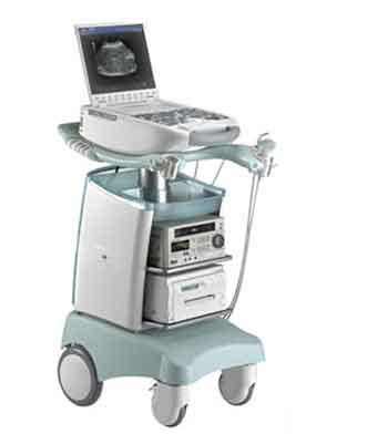
Figure 1: An example of ultrasound machine
(http://www.aadcoimaging.com)
During ultrasound scan, a gel is placed directly on the skin to help secure contact between the transducer and the skin. This will eliminate air pockets between the transducer and the skin that can block the sound waves from passing into the adjacent tissue. When the sound wave hit dense structures like the fetus and the uterus wall, the sound waves will be bounced back (reflected) to the transducer. The transducer then collects the reflected sound waves and translated it into a visual image by data processor. The image is then displayed on the screen.
Types of Ultrasound Imaging Mode
The following are the different types of images that can be formed using ultrasound:
- A-mode
A-mode or amplitude mode is the simplest type of ultrasound. It displays the amplitude of returning echoes as a function of depth, where x-axis represents the depth and y-axis represents the amplitude.
- B-mode
B-mode or brightness mode displays echoes as individual spots which are placed on the screen correspondent to their point of origin in the body. Differences in amplitudes of returning echoes are shown as different dot brightness.
B-mode imaging is the most widely used in ultrasound examination. Figure 2 shows an example of a B-mode image. Advancements in ultrasound technology made it possible to display the image in three-dimensional (3D) format. Figure 3 shows an example of a 3D image.
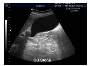
Figure 2: An example of B-mode image
(http://www.medison.ru/uzi/eho283.htm)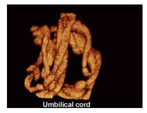
Figure 3: An example of 3D image
(http://www.medison.ru/uzi/eho211.htm) - Doppler ultrasound
Apart from B-mode, Doppler imaging is also widely use in medical examinations. Doppler ultrasound is a special type of ultrasound scan that evaluates blood flow through arteries and veins. The different speed and directions of blood flow detected are represented in colours for easier interpretation. Figure 4 shows an example of a Doppler image.
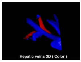
Figure 4: An example of Doppler image
(http://www.medison.ru/uzi/eho168.htm)
Advantages
Ultrasound has several advantages as compared to other medical imaging methods. The major advantage of ultrasound over computed tomography (CT) and conventional X-rays is that it does not use ionising radiation. Although MRI also does not use ionising radiation, MRI procedure requires patient sitting in a noisy and confined space. Moreover, MRI is much more expensive and not widely available as compared to ultrasound. In general, ultrasound scan is much faster than X-rays or other imaging methods.
Other advantages of ultrasound are as follows:
- It provides real-time imaging, making it a good tool for guiding medical procedures such as needle biopsies and fluid aspiration.
- It has no known long-term side effects and rarely causes any discomfort to the patient.
- Ultrasound scans are painless and easily tolerated by most patients. However, ultrasound procedure involving a transducer that is inserted into an opening of the body may produce minimal discomfort.
Disadvantages / Weakness
The disadvantage of ultrasound is more to the capability of the imaging tools itself. Thus, the term of “weakness” is more appropriate. The main weakness of ultrasound is the difficulty in imaging structures behind air and bones. Ultrasound waves are disrupted by air or gas; therefore ultrasound is not an ideal imaging technique for air-filled bowel or organs that obscured by the bowel. In most cases, barium exams, CT scan, and MRI are the methods of choice in such conditions. Ultrasound waves also have trouble to penetrate the bone. For example, imaging of the adult brain using ultrasound is very limited though improvements are being made in transcranial ultrasonography. Another drawback of ultrasound imaging is that it is relatively dependent on operator skills as compared to other imaging modalities.
Uses of Ultrasound in Medical
Ultrasound is useful in examining many of the body’s internal organs. It is also used during medical procedures. Some of the uses of ultrasound are as follows:
- Fetal ultrasound – to assess the fetal development in pregnancy and to check for any physical abnormalities.
- Abdominal ultrasound – to examine organs in the abdomen including liver, gallbladder, spleen, pancreas, and kidneys.
- Breast ultrasound – to look for breast cancer or other breast conditions.
- Echocardiogram – to look at the size, shape, and motion of the heart.
- Ophthalmic ultrasound – to examine ocular diseases such as iris melanoma.
- Guided interventional procedures – to assist in biopsies and drainage of fluid collections.
While Doppler ultrasound images, among others can help the doctors to see and evaluate:
- Blockages to blood flow such as clots.
- Narrowing of blood vessels.
- Tumors and congenital vascular malformations.
How Does the Procedure Work?
In ultrasound examination, a transducer is placed on directly and moved over the patient. Some types of ultrasound tests, however, need to be done with a transducer that is inserted into the body. This is done using transvaginal (endovaginal), transrectal (endorectal), and transesophageal (endoesophageal) transducers.
Risks and Side Effects
Diagnostic ultrasound is generally considered a safe imaging modality including imaging of the fetus. According to the British Medical Ultrasound Society, to date, there is no evidence that diagnostic ultrasound can cause any harm to humans, including the developing fetus. Similarly, World Health Organizations technical report series 875 (1998) stated that there is no published report of harmful biological effect. Nonetheless, Health Canada recommended that operators should be careful to limit exposure to critical organs.
Although there is no evidence that ultrasound could be harmful for the fetus, US Food and Drug Administration views use of ultrasound for making “keepsake fetal videos” to be an unapproved use of a medical device.
Summary
Ultrasound is widely used in medical to perform both diagnostic and to assist doctors to carry out certain procedures. B-mode image is the most common type used in ultrasound imaging while Doppler ultrasound is used to evaluate blood flow in the body. Despite its wide range of uses, the major drawback of ultrasound is its limitation in imaging structures behind bones and air. Unlike x-ray technique, ultrasound does not use ionising radiation. Instead, it uses sound wave to produce image. It is generally considered a safe imaging modality. To date, there is no evidence that diagnostic ultrasound has produced any harm to humans.
References
- Diagnostic Ultrasound: Physics and Equipment, ed. Peter Hoskins, Kevin Martin and Abigail Thrush. Cambridge University Press (2010)
- Guidelines for the safe use of diagnostic ultrasound equipment, The British Medical Ultrasound Society (2009)
- Guidelines for the safe use of diagnostic ultrasound, Health Canada, (2001)
- Training In Diagnostic Ultrasound: Essentials, Principles And Standards, WHO Technical Report Series 875 (1998)
- General Ultrasound (http://www.radiologyinfo.org – assessed: 10 October 2015)
| Last Reviewed | : | 04 March 2016 |
| Writer | : | Dr. Bidi bin Ab. Hamid |
| Accreditor | : | Bazli bin Sapiin |


