

Introduction
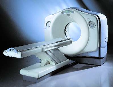
Figure 1: PET-CT machine
(Source: http://www.radiology-equipment.com/Detail.CFM?LineItemID=1638)
PET (Positron Emission Tomography) is an art imaging techniques used in the detection of metabolically active organ or tissues inside body. The concept of emission and transmission tomography was introduced by David E. Kuhl, Luke Chapman and Roy Edwards in the late 1950s. Their work later led to the design and construction of several tomographic instruments at the University of Pennsylvania, United State.
This imaging technique used primarily in oncology, neurology and cardiology. PET imaging very sensitive and able to detect small lesions in the body. The CT (Computed Tomography) component makes a PET-CT imaging modality capable of diagnosis various disease by using radiopharmaceutical f18 fluoro-deoxy-glucose (18F-FDG).
18F-FDG is a radioactive substance which is added to label with pharmaceuticals. The 18-FDG acts as a tracer and will emit gamma rays when administer into the patient’s body.
Nuclear Medicine Physician usually recommend patient undergoing PET-CT imaging in order to detect abnormal tissue or organ abnormality. This imaging technique is safe for patient and required small amount of F18-FDG injection.
PET-CT Detection Principle
PET-CT machine is composed of the PET which contains blocks of crystal to detect gamma ray from patients injected with F18-FDG.The CT component emit X-rays to performed patients anatomy and localizations.
After 18F-FDG is injected into the patient, it will be distributed to a specific organ. The organ would emit gamma rays and detected by PET to produce anatomical images and can give complete and accurate information about the cell, tissue and organ in fact able to detect abnormalities at an early stage.
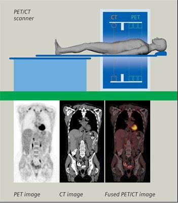
Figure 2: Two scan were perform separately and fused to produce PET-CT image
(Source: http://www.nepetimaging.com/pet-ct.html)
PET-CT In Medical Application
In nuclear medicine, PET-CT information is most frequently used for the detection of cancer and other diseases related secondary and it also can be used to monitor the effectiveness of treatment and restaging of cancer. In addition, it is also used to detect heart disease and brain disorders.
- Oncology (Cancer)
Brain tumours, head and neck tumours, ovarian cancer, thyroid carcinomas, colorectal cancer, pancreatic cancer, breast cancer, gastro-oesophageal cancer, bladder, liver cancer, lung cancer, renal and prostate cancer, cancer of uterus, testicular tumours, malignant melanoma, malignant lymphomas and musculoskeletal tumours.
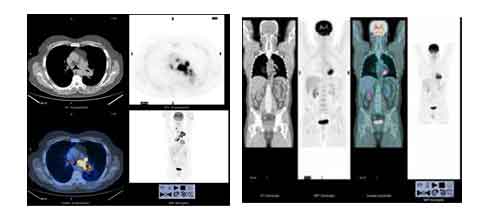
Figure 3: Sample of PET-CT imaging for Oncology diagnosis
(Source: Cook GJR, et al: Semin Nucl Med 1996;26:308-314) - Cardiology (Heart)
Coronary artery disease and myocardial infarction.
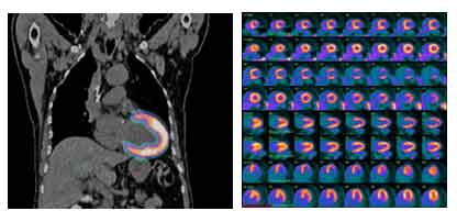
Figure 4: Sample of PET-CT imaging for Myocardial Infraction
(Source: http://www.radrounds.com and http://www.dicardiology.com) - Neurology (Brain)
Alzheimer’s disease, Epilepsy, Parkinson’s disease and Brain Tumor.
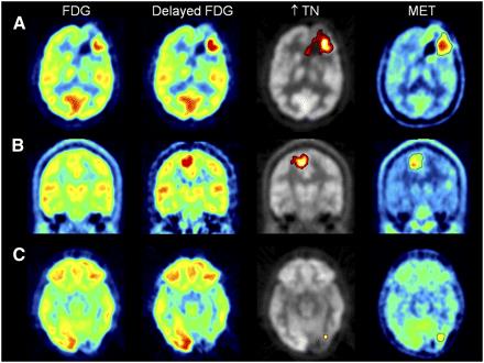
Figure 5: Sample of PET-CT imaging for brain abnormality
(Source: http://jnm.snmjournals.org/content/52/6/865/F3.expansion.html)
General Guideline Before PET-CT Procedures
Patients are advised to reduce strenuous activity involving the use of muscle directly at least two days before undergoing a PET scan. Patients also need to fast for a period of 4-6 hours before the PET-CT procedure, although the fasting period, the patient is allowed to drink plain water only.
For diabetics, patients are required to fast from 12 midnight on the day before the PET-CT procedure and required to take insulin injections before fasting. However, for other drugs the patient may be taking as usual.
General Guideline During PET-CT Procedure
On the day of the PET scan, patient was advised to arrive early to the scanning centre and bring together all the radiological reports. Patient was advised to come alone or accompanied by family member for radiation safety purpose.
Patient will be consult by Nuclear Medicine Physician before proceed with the PET-CT imaging.
Patients vital sign measurement shall be recorded before injected with F18-FDG.
Patients shall rest for one hour in a special room in order to make sure enough F18-FDG distribution to the patients before undergo PET-CT procedure.
Radiation Safety Guideline After PET Procedure
Patient can eat normal diet after the procedure, unless have received special instructions from Nuclear Medicine Physician.
Patients are encouraged to drink lots of water to get rid of 18F-FDG in the body through the urine. Patients are advised to stay at a distance of 1 meter from pregnant woman and children for good radiation safety practice. The F18-FDG will disappear within 7 hours to 24 hours from the body.
| Last Reviewed | : | 06 May 2016 |
| Writer/Translator | : | Rabi’atul Adawiyah bt. Mat Salleh |
| Accreditor | : | Mohamad Aminudin bin Said |


